Chapter 3 - CHICKEN ANATOMY AND PHYSIOLOGY
Chapter 3 - CHICKEN ANATOMY AND PHYSIOLOGY
Chapter 3 - CHICKEN ANATOMY AND PHYSIOLOGY
Create successful ePaper yourself
Turn your PDF publications into a flip-book with our unique Google optimized e-Paper software.
<strong>Chapter</strong> 3 - <strong>CHICKEN</strong> <strong>ANATOMY</strong> <strong>AND</strong> <strong>PHYSIOLOGY</strong><br />
Contents:<br />
Digestive system<br />
Respiratory system<br />
Skeletal system<br />
Muscle system<br />
Reproductive system - female<br />
Reproductive system - male<br />
Circulatory system<br />
Nervous system<br />
Excretory system<br />
Immune system<br />
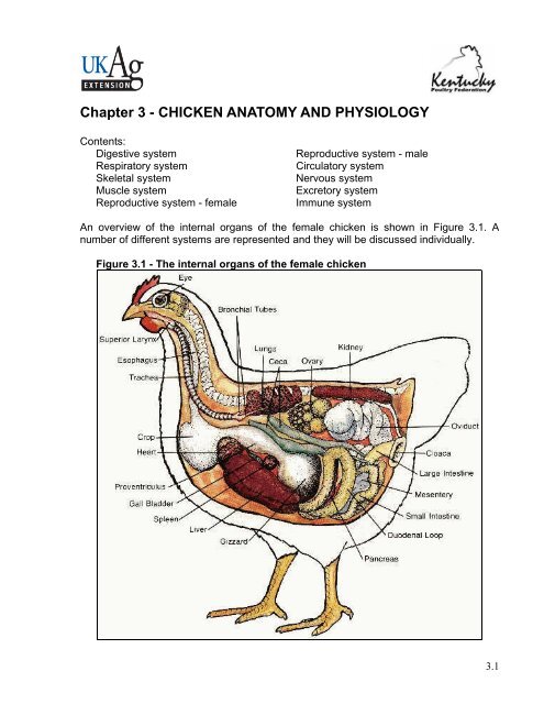
An overview of the internal organs of the female chicken is shown in Figure 3.1. A<br />
number of different systems are represented and they will be discussed individually.<br />
Figure 3.1 - The internal organs of the female chicken<br />
3.1
A. Digestive system<br />
The digestive system uses the nutrients in consumed feed for the maintenance of all the<br />
other systems of the chicken’s body. Ingested food is broken down to its basic<br />
components by mechanical and chemical means and these basic components are then<br />
absorbed and utilized throughout the body. A knowledge of the digestive process assists<br />
in understanding the nutritive requirements of chickens. In addition, knowing what’s<br />
‘normal’ can also help you recognize and take action when the digestive system goes<br />
awry. Frequent bouts with a particular digestive disorder, for example, may indicate a<br />
need for improved feeding or better sanitation.<br />
The avian digestive system begins at the mouth and ends at the cloaca and has several<br />
intervening organs in between (see Figure 3.2).<br />
3.2<br />
Figure 3.2 - The digestive tract of the chicken.<br />
• Beak / Mouth: Chicken’s obtain feed with the use of the beak. The feed then<br />
enters the digestive system via the mouth. The mouth contains glands that<br />
secrete saliva containing enzymes which begins the digestion of the feed<br />
consumed. The chicken does not have teeth to chew its feed. The tongue is used<br />
to push feed to the back of the mouth so that it can be swallowed. There are taste<br />
buds on the roof of the mouth and back of the tongue. The mouth is also very<br />
sensitive to temperature differences.
• Esophagus: The esophagus is a flexible tube that connects the mouth with the<br />
rest of the digestive tract. It carries food from the mouth to the crop and from the<br />
crop to the proventriculus.<br />
• Crop: The crop is an out-pocketing of the esophagus and is located just outside<br />
the body cavity in the neck region (see Figure 3.3). Consumed feed and water are<br />
stored in the crop until the remainder of the digestive tract is ready to receive<br />
more feed. When empty, or nearly empty, the crop sends hunger signals to the<br />
brain so that more feed is consumed. Although the mouth excretes the digestive<br />
enzyme amylase, very little, if any, digestion takes place in the crop – it is simply<br />
a temporary storage pouch that evolved for prey birds which need to move to the<br />
open to feed. They are able to consume relatively large quantities of food rapidly<br />
and then return to a more secure location to digest it. Occasionally the crop<br />
becomes impacted (crop impaction, also referred to as crop binding or<br />
pendulous crop). This may occur when feed is withheld for a period of time,<br />
causing chickens to eat too much too fast when the feed is returned. A crop may<br />
also become impacted in a chicken that is free-ranged on a pasture of tough,<br />
fibrous vegetation. With a crop impaction, even if a chicken continues to eat, the<br />
feed can not get past the impacted crop. The swollen crop may also cut off the<br />
windpipe, suffocating the chicken. Crop impaction is unlikely to occur in properly<br />
fed broilers or broiler breeders.<br />
Figure 3.3 - Photograph showing the location of the crop in a chicken. The crop is<br />
located just outside the body cavity in the neck region.<br />
• Proventriculus: The esophagus connects the crop to the proventriculus. The<br />
proventriculus (also known as the ‘true stomach’) is the glandular stomach<br />
3.3
3.4<br />
where digestion begins. As with our stomachs, hydrochloric acid and digestive<br />
enzymes (e.g., pepsin) are added to the feed here and digestion begins.<br />
• Gizzard / Ventriculus: The gizzard is a unique part of the avian digestive tract<br />
and is often referred to as the ‘mechanical stomach’. It is made up of two sets of<br />
strong muscles which act as the bird’s teeth. Consumed feed and the released<br />
digestive juices pass from the proventriculus to the gizzard for grinding, mixing,<br />
and mashing. Large poorly-soluble particles (such as small stones or grit) are<br />
retained in the gizzard until ground into tiny pieces by the action of the muscles<br />
and exposure to the acid and food particles. Broilers and broiler breeders fed only<br />
commercially prepared feed do not need grit. If, however, whole grains are fed<br />
without having access to grit, digestive efficiency will be impaired. When a<br />
chicken eats a small, sharp object such as a tack or staple, the object is likely to<br />
lodge in the gizzard, and due to the strong grinding motion of the gizzards<br />
muscles, may eventually pierce the gizzard wall. As a result, the chicken will grow<br />
thin and eventually die – a good reason to keep your poultry houses free of nails,<br />
glass shards, bits of wire and the like.<br />
• Small intestine: The small intestine is made up of the duodenum (also referred to<br />
as the duodenal loop) and the lower small intestine. The duodenum receives<br />
digestive enzymes and bicarbonate (to counter the hydrochloric acid from the<br />
proventriculus) from the pancreas and bile from the liver via the gall bladder.<br />
The digestive enzymes produced by the pancreas are primarily involved in protein<br />
digestion. The pancreas plays important roles in both the digestive and hormonal<br />
systems. It also secretes hormones into the blood system that are important in the<br />
regulation of blood sugar. Bile is a detergent that is important in the digestion of<br />
lipids and absorption of fat-soluble vitamins (vitamins A, D, E and K). The<br />
remainder of the digestion occurs in the duodenum and the released nutrients are<br />
absorbed mainly in the lower small intestine (jejunum and ileum). The lower<br />
small intestine is composed of two parts, the jejunum and ileum. The merkels<br />
diverticulum marks the end of the jejunum and the start of the ileum. Just prior to<br />
hatch, the yolk sac, which had been supplying nutrition during embryo<br />
development, is drawn into the navel cavity. The residual tiny sac is the merkels<br />
diverticulum. The yolk sac supplies feed and water to the newly hatched chick and<br />
is the reason that chicks can be shipped considerable distances (as in the postal<br />
service) without adverse effects. Omphalitis is a condition characterized by<br />
infected yolk sacs, often accompanied by unhealed navels in recently hatched<br />
chicks. It is infectious but not contagious. It is often associated with excessive<br />
humidity and marked contamination of the hatching eggs or incubator. The<br />
affected chicks usually appear normal until a few hours before death. Depression,<br />
drooping of the head, and huddling near the heat source usually are the only<br />
signs. The navel may be inflamed and fail to close, producing a wet spot on the<br />
abdomen; a scab may be present.<br />
• Ceca (plural form; singular = cecum): The ceca are two blind pouches at the<br />
junction of the small and large intestines. Re-absorption of water takes place in<br />
the ceca. Fermentation of coarse materials and production of the eight B vitamins<br />
(Thiamine, riboflavin, niacin, pantothenic acid, pyridoxine, biotin, folic acid and<br />
vitamin B12) also occur in the ceca, but because the ceca are located near the
end of the digestive tract there is minimal absorption of any nutrients released.<br />
The ceca empty their contents two or three times a day, producing pasty<br />
droppings that often smell worse than regular droppings and often mustard to<br />
dark brown in color. The frequency of cecal droppings, as well as their<br />
appearance among regular droppings, tells you the chicken’s digestive tract is<br />
functionally normally.<br />
• Large intestine (also known as the colon): Despite the name, the large<br />
intestine is actually shorter than the small intestine. The large intestine is where<br />
the last of the water re-absorption occurs.<br />
• Cloaca: In the cloaca there is a mixing of the digestive wastes together with<br />
wastes from the urinary system (urates). Fecal material is usually voided as<br />
digestive waste with white uric acid crystals on the outer surface (i.e., chickens do<br />
not urinate/pee). The reproductive tract also exits through this area (e.g., eggs or<br />
sperm).<br />
Both the small and large intestine are normally populated by beneficial bacteria, referred<br />
to as microflora (‘micro’ meaning small and ‘flora’ meaning plants). Microflora aid in<br />
digestion and enhance immunity by guarding their territory (i.e., the digestive tract)<br />
against invading microbes. Intestinal disease normally occurs when the balance of<br />
microflora is upset or the normal microflora is overrun by too many foreign organisms.<br />
The result is enteritis or inflammation of the intestines, producing symptoms that include<br />
diarrhea, increased thirst, dehydration, loss of appetite, weakness, and weight loss or<br />
slow growth.<br />
Chicken Feces<br />
The color and texture of chicken fecal material can indicate the health status of the<br />
chicken’s digestive tract. The white pasty material that commonly coats chicken fecal<br />
material is uric acid, the avian form of urine, and is normal (see Figure 3.4).<br />
Figure 3.4 - Normal chicken manure<br />
3.5
Some of the possible abnormal color and texture changes that can occur, together with<br />
possible causes, are shown below. These are just possible causes and not a definite<br />
cause. If you notice any abnormalities, notify your service person as soon as possible.<br />
3.6<br />
Appearance of Feces<br />
• Droppings with blood = coccidiosis<br />
• Greenish droppings = late stages of worms (or has eaten a lot of green<br />
vegetables if free-ranged)<br />
• White, milky runny droppings = worms, coccidiosis, Gumboro disease<br />
(Infectious Bursal Disease)<br />
• Brown runny droppings = E. coli infection<br />
• Clear or watery runny droppings = stress, Infectious Bronchitis<br />
• Yellow & foamy droppings = coccidiosis<br />
• Grayish white & running continuously = vent gleet (a chronic disease of the<br />
cloaca of domestic birds)<br />
B. Respiratory system<br />
The respiratory system is involved in the absorption of oxygen, release of carbon<br />
dioxide, release of heat (temperature regulation), detoxification of certain chemicals,<br />
rapid adjustments of acid-base balance, and vocalization. While the function of the avian<br />
respiratory system is comparable to that of mammals, the two are quite different<br />
anatomically. Birds don’t breathe the same way mammals do. Like mammals, birds have<br />
two symmetrical lungs that are connected to a trachea (windpipe). But here the similarity<br />
ends. Mammalian lungs contain many bronchi (tubes), which lead to small sacs called<br />
alveoli. Because alveoli have only one opening, air can flow into and out of them, but it<br />
can not flow through them to the outside of a lung. In comparison, the avian lung has<br />
parabronchi which are continuous tubes allowing air to pass through the lung in one<br />
direction. They are laced with blood capillaries and it is here that gas exchange occurs.<br />
The trachea divides into two smaller tubes called bronchi (plural form; singular =<br />
bronchus). In some respiratory diseases tracheal ‘plugs’ are often formed and they<br />
physically block the respiratory tract at the junction of the bronchi. As a result, the<br />
chickens suffocate. Excessive dust in the air is also believed to result in the formation of<br />
caseous tracheal plugs and adversely affect the health of the chickens.<br />
The avian respiratory tract (Figures 3.5 and 3.6) starts with the glottis which closes<br />
when feed is passing down the throat so that feed does not enter the lungs. The trachea<br />
is made up of cartilaginous rings that prevent its collapse from the negative pressure<br />
caused by inspiration of air. The syrinx is the voice box. The chicken ‘voice’ is produced<br />
by air pressure on a sound valve and modified by muscle tension. It is not possible to<br />
remove the syrinx to prevent roosters from crowing. Both roosters and hens are able to<br />
‘crow.’ The reason hens don’t normally crow is because they ‘don’t feel like it’ due to<br />
female hormone effects and the absence of sufficient levels of the male hormone. When<br />
the ovaries become diseased and the level of female hormones decrease, many hens<br />
will start to show male characteristics, including crowing.
Figure 3.5 – Illustration showing<br />
the parts of the avian<br />
respiratory tract.<br />
Figure 3.6 - Illustration showing the location of<br />
the avian air sacs.<br />
The lungs are relatively small and do not expand. Instead, they are firmly attached to<br />
ribs. Birds have an incomplete diaphragm and the arrangements of the chest<br />
musculature and the sternum do not lend themselves to expansion in the same way that<br />
the chest of mammals does. Consequently they can’t inflate and deflate lungs in the<br />
same way as mammals do. Instead, birds pass air through the lungs by means of air<br />
sacs, a uniquely avian anatomical feature. The air sacs are balloon-like structures at the<br />
‘ends’ of the airway system. In the chicken there are nine such sacs: an unpaired one in<br />
the cervical region; two interclavicular air sacs, two abdominal air sacs, two anterior<br />
thoracic air sacs and two posterior thoracic air sacs (see Figure 3.7). The avian<br />
respiratory system is described as non-tidal. The mammalian respiratory system, in<br />
contrast, is tidal.<br />
Figure 3.7 - Dorsal view of the air sac locations in chickens<br />
3.7
The key to the avian respiratory system is that distention and compression of the air<br />
sacs, not the lungs, moves air in and out. At any given moment air may be flowing into<br />
and out of the lung and being ‘parked’ in the air sacs (see Figure 3.8). The lungs are stiff<br />
and fixed, not at all like the distensible lungs of mammals. The air sacs act as ‘bellow’s<br />
to suck air in and blow it out and also to hold part of the total volume. The air sacs fill a<br />
large proportion of the chest and abdominal cavity of birds, and also connect to the air<br />
spaces in the bones.<br />
3.8<br />
Figure 3.8 - The flow of air through the avian respiratory system.<br />
1. On first inhalation, air flows through the trachea & bronchi, primarily into the<br />
posterior (rear) air sacs<br />
2. On exhalation, air moves from the posterior air sacs into the lungs<br />
3. With the second inhalation, air moves from the lungs into the anterior (front) air<br />
sacs<br />
4. With the second exhalation, air moves from the anterior air sacs back into the<br />
trachea and then out<br />
Figure 3.9 - Diagram showing movement of sternum and ribs during respiration<br />
A. Inspiration; B. Expiration; C. Sternum (keel)
Since birds do not have a diaphragm, they depend on the movement of the sternum<br />
(keel) and rib cage in order to breathe (see Figure 3.9). Holding a bird too tight will<br />
restrict movement of the rib cage and suffocate the bird. This often happens when young<br />
children hold baby chicks.<br />
With each breath, the chicken’s respiratory tract is exposed to the inside environment of<br />
a poultry house. Poor environments normally do not cause disease directly but they do<br />
reduce chickens’ defenses, making them more susceptible to existing viruses and<br />
pathogens.<br />
The air of poultry houses can contain aerosol particles or ‘dust’ originating from the<br />
floor litter, feed, dried manure, and the skin and feathers of the chickens. These aerosol<br />
particles can have a range of adverse effects on poultry. They act as an irritant to the<br />
respiratory system and coughing is a physiological response designed to remove them.<br />
Excessive coughing lowers the chicken’s resistance to disease. Aerosol particles often<br />
collect inside the chicken and can increase carcass condemnation at the processing<br />
plant.<br />
The chicken’s respiratory tract is normally equipped with defense mechanisms to prevent<br />
or limit infection by airborne disease agents; to remove inhaled particles; and to keep the<br />
airways clean. Chicken health is affected by the function of three defensive elements:<br />
the cilia; the mucus secretions; and the presence of scavenging cells which consume<br />
bacteria.<br />
Cilia are tiny hair-like structures in the trachea. Cilia are responsible for propelling the<br />
entrapped particles for disposal. Mucus is produced in the trachea. Mucus secretion and<br />
movement of cilia are well developed in chickens. The consistency of the mucus<br />
produced is important for the efficiency of the ciliary activity. Cilia cannot function when<br />
the mucus is too thick.<br />
Scavenging cells in the lungs actively ‘scavenge’ inhaled particles and bacteria that<br />
gain entrance to the lower respiratory tract. These cells consume bacteria and kill them,<br />
thus preventing their further spread.<br />
It is the integrated function of cilia, mucus and scavenging cells that keeps broiler<br />
airways free of disease-producing organisms. The impairment of even one of these<br />
components permits an accumulation of disease agents in the respiratory tract and may<br />
result in disease.<br />
Gases are generated from decomposing poultry waste; emissions from the chickens;<br />
and from improperly maintained or installed equipment, such as gas burners. Harmful<br />
gases most often found in poultry housing are ammonia (NH3) and carbon dioxide (CO2).<br />
Research has shown that as little as 10 ppm ammonia will cause excessive mucus<br />
production and damage to the cilia. Research has also revealed that ammonia levels of<br />
10-40 ppm reduce the clearance of E. coli from air sacs, lungs, and tracheas in chickens.<br />
3.9
C. Skeletal system<br />
Aside from the obvious role of structural support, the skeletal system (see Figure 3.10)<br />
has two additional functions: respiration and calcium transport.<br />
The skeletal system of the bird is compact and lightweight, yet strong. The tail and neck<br />
vertebrae are movable, but the body vertebrae are fused together to give the body<br />
sufficient strength to support the wings. There are two special types of bones which<br />
make up the bird’s skeletal system: the pneumatic and medullary bones.<br />
3.10<br />
Figure 3.10 - Illustration of the chicken's skeleton.
The pneumatic bones are important to the chicken for respiration. They are hollow<br />
bones which are connected to the chicken’s respiratory system and are important for the<br />
chicken to breathe. Examples of pneumatic bones are the skull, humerus, clavicle, keel<br />
(sternum), pelvic girdle, and the lumbar and sacral vertebrae.<br />
The medullary bones are an important source of calcium for the laying hen. Calcium is<br />
the primary component of egg shell and a hen mobilizes 47% of her body calcium to<br />
make the egg shell. Examples of medullary bones are the tibia, femur, pubic bones, ribs,<br />
ulna, toes, and scapula.<br />
D. Muscle system<br />
There are three types of muscles in the chicken’s body: smooth, cardiac, and skeletal.<br />
Smooth muscle is controlled by the autonomic nervous system (ANS) and is found in<br />
the blood vessels, gizzard, intestines and organs. The cardiac muscle is the specialized<br />
muscle of the heart. The skeletal muscle is the type of muscle responsible for the<br />
shape of the bird and for its voluntary movement. This is the muscle type that makes up<br />
the edible portions of the carcass. The most valuable skeletal muscles in a poultry<br />
carcass are the breast, thigh and leg.<br />
The breast meat is referred to as ‘white meat’. White meat is ‘white’ because of a lower<br />
level of exercise for these muscles. The thigh and leg meat are referred to as ‘dark<br />
meat.’ Dark meat is ‘dark’ because the muscles are used for sustained activity – in the<br />
case of a chicken, chiefly walking. The dark color comes from a chemical compound in<br />
the muscle called myoglobin, which plays a key role in oxygen transport. White muscle,<br />
in contrast, is suitable only for short, ineffectual bursts of activity such as, for chickens,<br />
flying. That's why the chicken's leg meat and thigh meat are dark and its breast meat<br />
(which makes up the primary flight muscles) is white. Other species of poultry more<br />
capable of flight (such as ducks, geese, and guinea fowl) have dark meat throughout.<br />
The main objective of the broiler industry is the production of SALEABLE chicken meat.<br />
To this end, it is important to limit to a minimum the number of condemnations at the<br />
processing plant and to maximize meat yield. Production of a quality meat product from<br />
a live broiler chicken involves a series of efficiently-performed, specific tasks carried out<br />
in a sanitary manner. Before broilers can be processed they must be raised to market<br />
age, caught, cooped, transported and held; then unloaded at the processing plant. Inside<br />
the processing plant, broilers are hung on shackles, stunned, bled, de-feathered,<br />
eviscerated, inspected, chilled, graded, and packaged. Because of the complexity of<br />
production and processing procedures, several factors may reduce carcass yield and<br />
adversely affect carcass grade. Defects resulting in carcass downgrading and product<br />
losses are caused by disease, damage to the chicken before slaughter, or maladjusted<br />
equipment and manual errors during processing.<br />
Good live haul procedures can reduce bruising and the number of DOAs (dead on<br />
arrivals). Broilers that are transported long distances (6+ hours) have fewer bruises due<br />
to confinement, and the fatigue from traveling makes them less active during slaughter.<br />
However, the stress associated with transporting slows digestive tract clearance and<br />
increases the live shrink (live weight loss).<br />
3.11
With regard to meat quality, the poultry industry has seen an increase in the occurrence<br />
of Pale Soft and Exudative (PSE) meat over the past several years. PSE meat is the<br />
result of accelerated postmortem glycolysis, which results in a rapid pH decline while<br />
carcass temperatures are still high. Processors are concerned with the appearance of<br />
PSE meat in fresh tray packs as the pale color can affect color uniformity within the<br />
package. Although pale meat is still healthy, it is less pleasing to the customer. All<br />
poultry meat looses moisture, but pale meat looses slightly more. Pale meat is estimated<br />
to result in an annual loss of about $200 million for the U.S. broiler industry.<br />
Another meat quality concern is Green Muscle Disease or Deep Pectoral Myopathy<br />
(see Figure 3.11), which is the necrosis of the Pectoralis Minor (tender) muscle of<br />
poultry. Necrotic tenders appear yellowish-green in color. Green Muscle Disease results<br />
from vigorous activity of both major and minor pectoralis muscles, but only the tenders<br />
are afflicted. During contraction, muscles normally swell with increased blood flow to<br />
supply needed oxygen and nutrients. Compared to other muscles, the tenders have a<br />
more rigid muscle cover and are confined to a tight space within the body such that they<br />
can’t expand to accommodate this increased blood flow. The net result of the muscle<br />
being confined and compressed is self strangulation, suffocation, and eventually<br />
necrosis.<br />
Figure 3.11 - Chicken with green muscle disease<br />
The incidence of Green Muscle Disease increases with higher market weight in broilers,<br />
and more cases are reported in higher yielding crosses and in males. Increased broiler<br />
activity induced by such factors as feed or water outages, lighting programs, catching<br />
and live haul, and even excessive noise, may result in an increased incidence of Green<br />
Muscle Disease. The increased activity associated with free-range broiler production has<br />
resulted in an increase incidence of Green Muscle Disease in this industry.<br />
3.12
E. Reproductive system - female<br />
The avian reproductive system is very different from that of mammals and is better<br />
suited to the peculiar hazards of being a bird. The biggest problem with being a bird is<br />
that everyone is trying to eat you. While most mammals (especially primates) have<br />
adopted a strategy of having relatively few offspring and devoting a fair amount of<br />
parental energy and time to caring for those few, most birds (with some notable<br />
exceptions) have gone the other way: they produce lots of offspring, give them some<br />
minimal amount of raising – in some cases, none – then toss them to the winds, literally<br />
and figuratively. The parts of an incubated egg are shown in Figure 3.12.<br />
The female reproductive system of the chicken (Figure 3.13) is divided into two main<br />
parts: the ovary and the oviduct. In the majority of avian species, including chickens, only<br />
the left ovary and oviduct are functional. Although the right is present embryologically, it<br />
regresses during development and is vestigial in the adult bird.<br />
Figure 3.12 - Interior views of an incubated chicken's egg.<br />
The ovary is a cluster of developing yolks or ova and is located midway between the<br />
neck and tail of the bird, attached to the back. The ovary is fully formed although very<br />
small when the female chick is hatched. It is made up of 13,000 – 14,000 ova which<br />
grow by the addition of yolk fluid. Each ovum (singular of ova) starts out as a single cell<br />
surrounded by a vitelline membrane. As the ovum develops, yolk is added. The color of<br />
the yolk comes from fat soluble pigments called xanthophylls contained in the hen’s<br />
diet. At hatch, chicks (like codfish and mammals) are equipped with tens of thousands of<br />
potential eggs which theoretically could be laid, but (as with the oocytes of mammals)<br />
most of them never develop to the point of ovulation. No new ova are produced once the<br />
chick has hatched.<br />
Ovulation is the release of the mature ovum from the ovary into the second part of the<br />
female reproductive system, the oviduct. The ovum, which is enclosed in a sac, ruptures<br />
along the suture line or stigma. This release of the ova occurs 30-75 minutes after the<br />
previous egg has been laid.<br />
3.13
Figure 3.13 - Photograph of a hen's reproductive system.<br />
The second major part of the female chicken’s reproductive system is the oviduct. The<br />
oviduct is a long convoluted tube (25-27 inches long when fully developed) which is<br />
divided into five major sections. They are the infundibulum or funnel, magnum, isthmus,<br />
shell gland, and vagina.<br />
The first part of the oviduct, the infundibulum or funnel, is 3-4 inches long, and it<br />
engulfs the ovum released from the ovary. The ovum or yolk remains in the infundibulum<br />
15-18 minutes. The infundibulum also serves as a reservoir for spermatozoa so that<br />
fertilization can take place.<br />
The next section of the oviduct is the magnum which is 13 inches long and is the largest<br />
section of the oviduct as its name implies (from the Latin word for ‘large’). The ovum or<br />
yolk remains here 3 hours during which time the thick white or albumen is added.<br />
The third section of the oviduct is the isthmus which is 4 inches long. The ‘egg’ remains<br />
here for 75 minutes. The isthmus, as its name implies, is slightly constricted (The term<br />
3.14
‘isthmus’ refers to a narrow band of tissue connecting two larger parts of an anatomical<br />
structure). The isthmus is where the inner and outer shell membranes are added.<br />
The next section of the oviduct is the shell gland or uterus. The shell gland is 4-5 inches<br />
long, and the ‘egg’ remains here for 20 plus hours. As its name implies, the shell is<br />
placed on the egg here. The shell is largely made up of calcium carbonate. The hen<br />
mobilizes 47% of her body calcium from her bones to make the egg shell, with the diet<br />
providing the remainder of the required calcium. Pigment deposition is also done in the<br />
shell gland.<br />
The last part of the oviduct is the vagina which is about 4-5 inches long and does not<br />
really play a part in egg formation. The vagina is made of muscle which helps push the<br />
egg out of the hen’s body. There are also glands located in the vagina where<br />
spermatozoa are stored. The bloom or cuticle is also added to the egg in the vagina<br />
prior to oviposition (the laying of the fully formed egg).<br />
Near the junction of the vagina and the shell gland, there are deep glands lined with<br />
simple columnar epithelium. These are the sperm host glands, so called because they<br />
can store sperm for long periods of time (10 days to 2 weeks!). When an egg is laid,<br />
some of these sperm can be squeezed out of the glands into the lumen of the tract, so<br />
that they can migrate farther up the oviduct to fertilize another egg. This is one of the<br />
really remarkable things about birds; the sperm remain viable at body temperature.<br />
Birds lay eggs in clutches. A clutch consists of one or more eggs, followed by a rest<br />
period of about a day, then another egg or set of eggs. Clutch size is species-specific<br />
and is held within fairly tight limits. Clutch size, as well as the numbers of clutches laid in<br />
a breeding season, will vary with species, but the principle is the same.<br />
In hens, ovulation usually occurs in the morning and under normal daylight conditions,<br />
almost never after 3:00 PM. The total time to form a new egg is about 25-26 hours. This<br />
includes about 3½ hours to make the albumen, 1½ hours for the shell membranes, and<br />
about 20 hours for the shell itself.<br />
Ovulation for the next egg of a clutch occurs within an hour of laying the previous egg,<br />
and so that each day the hen gets later and later in her timing; she "runs behind," like a<br />
clock that is improperly adjusted. Eventually she gets so far behind schedule that she<br />
would have to ovulate later than 3:00 PM. Since hens do not typically ovulate after 3:00<br />
PM, the next ovulation is delayed and egg laying is interrupted. This delay results in the<br />
break between clutches and the cycle repeats itself a day or so later.<br />
Occasionally, a hen will produce double-yolked eggs. This phenomenon occurs<br />
primarily due to the age of the hen, but can also be related to genetics. Young hens<br />
sometimes release two follicles from the ovary in quick succession. The highly active<br />
ovary due to high activity of reproductive hormones in peak egg production can also be a<br />
factor. Double-yolked eggs are larger in size than single yolk eggs. Double-yolked eggs<br />
are not suitable for hatching. There is typically not enough nutrients and space available<br />
for two chicks to develop to hatch. It has happened, but it is rare.<br />
3.15
Occasionally an egg contains more than two yolks. The Guinness Book of Records lists<br />
the world's largest chicken egg (with a diameter of 9 inches or 22.5 cm) as having five<br />
yolks and the heaviest egg (1 pound or 0.45 kg) as having a double yolk and a double<br />
shell.<br />
It is rare, but not unusual, for a young hen to produce an egg with no yolk at all.<br />
Yolkless eggs are usually formed when a bit of tissue is sloughed off the ovary or<br />
oviduct. This tissue stimulates the secreting glands of the different parts of the oviduct<br />
and a yolkless egg results.<br />
Things occasionally go wrong when an egg shell is being developed. The most obvious<br />
relates to shell texture (Figure 3.14). Occasionally the shell becomes damaged while still<br />
in the shell gland and is repaired prior to being laid. This results in what is known as a<br />
‘body check.’ Occasionally there will be ‘thin spots’ in the shell or ‘ridges’ will form.<br />
The shells of such eggs, though not cracked, are weaker than ‘normal’ eggs and should<br />
not be used as hatching eggs. A second category of problems is abnormal shape<br />
(Figure 3.15). To be considered a hatching egg, the egg should be the typical ‘egg<br />
shape.’ Abnormally shaped eggs should not be used as hatching eggs. In many cases it<br />
is not clear which is the large end (and eggs should be incubated large end up) or they<br />
may not properly fit in the egg trays.<br />
Occasionally an egg will be laid without a shell. It feels like a water balloon. The<br />
membrane was placed on the yolk and white, but it somehow slipped past the "shell<br />
mechanism" and the shell wasn't deposited. The occurrence of the occasional shell-less<br />
egg is not necessarily an indication of any disease problem. If the incidence increases,<br />
however, a veterinarian should be consulted.<br />
Even rarer is an egg within an egg. This occurs when an egg that is nearly ready to be<br />
laid reverses direction and gets a new layer of albumen covered by a second shell.<br />
Sometimes the reversed egg joins up with the next egg and the two are encased<br />
together within a new shell. Double shelled eggs are so rare that no one knows exactly<br />
why or how they happen.<br />
Figure 3.14 - Examples of poor exterior egg quality related to shell texture.<br />
A. Body check<br />
B. Thin spot<br />
C. Ridges<br />
3.16
Figure 3.15 - Examples of abnormally shaped eggs.<br />
A. Pear-shaped egg<br />
B. Football-shaped egg<br />
F. Reproductive system - male<br />
The avian male reproductive system (Figures 3.16 and 3.17) is all inside the bird – unlike<br />
mammals which have the testes outside of the body. The male chicken possesses two<br />
testes, along the back, near the anterior ends of the kidneys. They are elliptical shaped<br />
and light yellow in color. Each ductus deferens opens into a small bump, or papilla,<br />
which is on the dorsal wall of the cloaca. The papillae serve as the copulatory organ. The<br />
incorrectly named, "rudimentary copulatory organ" is located on the medial ventral<br />
portion of the cloaca and is used to classify the sex of baby chicks.<br />
Figure 3.16 - Diagram of the<br />
avian male reproductive tract<br />
Figure 3.17 - Photo of a male<br />
chicken’s reproductive tract<br />
The main goal of broiler breeder management is to produce hatching eggs. However,<br />
the only good hatching egg is a fertilized egg. Fertility, the percentage of eggs laid that<br />
are fertilized, is very important in hatching egg production. If an egg is not fertilized, then,<br />
3.17
of course, it will not contain an embryo and will not hatch. Simply put, "Hatchability can<br />
never be better than fertility."<br />
Fertility is affected by both the male and the female, and both tend to decline as the<br />
chickens age. Flock fertility is dependent on the reproductive status of the chickens (i.e.,<br />
level of egg and semen production) combined with the chickens’ interest and capability<br />
of mating. The fertility of a broiler breeder flock usually increases from a low of 65-75%<br />
at the start of lay (23-24 weeks of age) and peaks at 95-98% at 35-37 weeks of age.<br />
Between 40-45 weeks of age fertility declines and the older the birds get the faster the<br />
decline in fertility. From the female side, the decline in fertility is believed to be due to<br />
faster release of sperm from the sperm storage tubules. As a result, after 40 weeks of<br />
age the breeder hen needs more frequent mating to sustain high fertility. From the male<br />
side it is presumed that there is a decrease in sperm quality as the rooster ages, as well<br />
as a decrease in mating activity. There is also believed to be an increase in early embryo<br />
death in the second half of the reproduction cycle. These early deaths often appear as<br />
‘clears’ and may be mistaken for infertiles.<br />
Walking through the supermarket, it is often possible to find capons for sale. While it is<br />
obvious from the shape of the packaging that it is a bird of some kind, there is often no<br />
indication in the labeling of what exactly a capon is. An informal survey of 4-Hers with<br />
poultry projects and university students very few knew what a capon is. In caponization,<br />
the surgical castration of male chickens, the testes of the male chicken are completely<br />
removed. As a result, the cockerel fails to develop certain male characteristics or tends<br />
to lose them if they are developed. Capons are usually quiet and docile, lacking a<br />
cockerel's disposition to fight. The comb and wattles cease growing after castration, so<br />
the head of a capon looks small. The hackle, tail and saddle feathers grow unusually<br />
long.<br />
Removal of the testes, and thus elimination of the male sex hormones they produce,<br />
reduces the male sex instinct and changes their behavior. They will become more docile<br />
and less active. Energy that is normally expended in fighting, courting behavior, and<br />
territorial protection is greatly reduced, allowing more efficient conversion of feed into<br />
growth, fat deposition and improved meat quality.<br />
Caponizing produces a unique type of poultry meat grown for a specialized market. The<br />
meat of uncastrated cockerels tends to become rather coarse, stringy, and tough as the<br />
chickens age. This is not the case with the capon. Caponized males grow more slowly<br />
than normal male chickens and accumulate more body fat. The concentration of fat in<br />
both the light and dark meat of capons is greater than that of intact males. It is claimed<br />
that the capon meat is more tender, juicier, and more flavorful than regular chicken.<br />
Any breed of chicken can be caponized. Over the past 100 years breeds that were<br />
particularly favored for capon production included Jersey Giants, Brahmas, Orpingtons,<br />
Cornish, Plymouth Rocks, and Cochins. Today commercially grown capons are<br />
produced using the Cornish x Plymouth Rock cross typically used by the commercial<br />
broiler industry. Male birds are typically caponized at two to four weeks of age. The<br />
testes of a male chicken are located within the abdominal cavity. A good caponizer can<br />
operate on about 200 birds per hour. Commercially grown capons are marketed at 15 to<br />
3.18
18 weeks of age. The goal is a capon weighing six to eight pounds at packaging (9 to 11<br />
pounds live weight).<br />
Today, relatively few capons are marketed commercially in the United States. It is<br />
estimated that around a million capons are produced annually. This is in contrast with<br />
the 8 billion broilers produced each year. Today commercial capon production in the<br />
United States is now limited to a single producer, Wapsie Produce Company in Iowa.<br />
G. Circulatory system<br />
The avian circulatory system consists of a heart plus vessels that transport nutrients,<br />
oxygen and carbon dioxide, waste products, hormones, and heat. The vascular system<br />
of birds differs from that of mammals in that there is an additional renal portal system.<br />
Venous blood from the legs flows directly to the kidneys and further to the posterior vena<br />
cava.<br />
Figure 3.18 - Diagram of the avian heart. Birds, like mammals, have a 4-chambered<br />
heart (Figure 3.16), with complete<br />
separation of oxygenated and deoxygenated<br />
blood. The right ventricle<br />
pumps blood to the lungs, while the left<br />
ventricle pumps blood to the rest of the<br />
body. Because the left ventricle must<br />
generate greater pressure to pump blood<br />
throughout the body (in contrast to the right<br />
ventricle that pumps blood to the lungs),<br />
the walls of the left ventricle are much<br />
thicker & more muscular.<br />
Birds tend to have larger hearts than mammals (relative to body size and mass). The<br />
relatively large hearts of birds may be necessary to meet the high metabolic demands of<br />
flight. Among birds, smaller birds have relatively larger hearts (again relative to body<br />
mass) than larger birds. Hummingbirds, for example, have the largest hearts (relative to<br />
body mass) of all birds, probably because hovering takes so much energy.<br />
Avian hearts also tend to pump more blood per unit time than mammalian hearts. In<br />
other words, cardiac output (amount of blood pumped per minute) for birds is typically<br />
greater than that for mammals of the same body mass. Cardiac output is influenced by<br />
both heart rate (beats per minute) and stroke volume (blood pumped with each beat).<br />
'Active' birds increase cardiac output primarily by increasing heart rate.<br />
The red and white blood cells are formed in the spleen. A bird’s red blood cells are<br />
unique in that they are nucleated (there is a nucleus) whereas a mammal’s are not.<br />
Significant diseases of the cardiovascular system in poultry meat inspection include<br />
pericarditis and ascites. Pericarditis is an inflammation of the fibroserous membranous<br />
sac called pericardium, which encloses and lubricates the heart. Ascites is an increase<br />
of fluid in one or more of the abdominal spaces.<br />
3.19
Broilers subject to high environmental temperatures exhibit many behavioral changes<br />
which allow them to re-establish heat balance with their surrounds. They tend to rest<br />
more or will position themselves close to walls or waterers. They spread their wings<br />
away from the body to promote cooling by reducing body insulation. Within the bird,<br />
blood is diverted from certain internal body organs such as the liver, kidneys and<br />
intestines to dilated blood vessels of the peripheral tissue (i.e., skin) in order to facilitate<br />
heat loss.<br />
As ambient air temperatures increase past 85°F, chickens respond by trying to loose<br />
more heat through evaporative cooling which is accomplished by panting. However, this<br />
process also generates more heat through the muscular activity involved in panting.<br />
Normally blood pH is controlled by the lungs and kidneys along with the various buffer<br />
systems which prevent rapid changes in the pH. As the respiratory rate increases in heat<br />
stressed broilers, however, there is also a corresponding decrease in the levels of blood<br />
carbon dioxide. Respiratory alkalosis (i.e., elevated blood pH) results as the blood<br />
acid-base balance is disrupted. This produces a decrease in blood calcium and<br />
bicarbonate which are necessary for the production of strong egg shells. Thus, the<br />
ultimate problem with broiler breeders is a production of thin-shelled eggs produced by<br />
laying hens. As for growing chickens, heat stress affects them by depressing weight gain<br />
mainly because feed intake is depressed.<br />
H. Nervous system<br />
The nervous system is divided into two main parts which are the central nervous<br />
system (CNS) and the autonomic nervous system (ANS). The CNS is responsible for<br />
the voluntary actions of the body such as movement of flight, and the ANS is responsible<br />
for the coordination of involuntary actions of the organs, intestines, blood vessels and<br />
glands. The primary function of the nervous system is to integrate the functions of the<br />
body.<br />
Electrical stunning, using a water bath stunner, is the most common method employed<br />
to slaughter poultry under commercial conditions. The purpose of electrical stunning is to<br />
induce insensibility in order to perform humane neck cutting and to avoid recovery of<br />
consciousness and wing flapping during bleeding. The basic principle involved in<br />
electrical stunning is that an electrical current is passed through the brain to induce<br />
epilepsy. The occurrence of grand mal epilepsy is considered to be an indicator of the<br />
state of unconsciousness (based on the human analogy).<br />
Controlled Atmosphere Killing (CAK) is an alternative method for slaughtering poultry.<br />
It involves placing the chickens in a container in which the atmosphere lacks oxygen and<br />
is made up of argon and/or nitrogen and/or CO2. The result is that the chickens loose<br />
consciousness. Argon and nitrogen are important components of a painless gassing<br />
process as using CO2 seems to cause distress. The respiratory system has CO2<br />
detectors and high levels of CO2 will rest in a fighting response in chickens – typically<br />
wing flapping – which can result in increased wing damage. There are no such receptors<br />
for argon or nitrogen.<br />
3.20
I. Excretory system<br />
The main organ of the excretory system is the kidney. The functional units of kidneys<br />
are nephrons. The functions of the excretory system are to excrete water and metabolic<br />
wastes and to regulate the acid-base balance of the bird’s body.<br />
The two kidneys of the domestic fowl each generally with three lobes are found<br />
immediately behind the lungs on each side of the vertebral column and closely<br />
associated with it (Figure 3.19). They are brownish in color and their consistency is such<br />
that they are easily damaged during their removal. The kidneys are normally left in when<br />
a broiler carcass is processed.<br />
Figure 3.19 - Photograph indicating location of the kidneys in back of a chicken.<br />
Each kidney is divided into three distinct lobes. Chickens do not have a bladder. Urine<br />
passes from the kidney, through the ureters, which then empty into the cloaca. Chicken<br />
urine consists of yellowish fluid with a white pasty substance largely made of uric acid.<br />
Uric acid is the main product of nitrogen metabolism in birds and is not water<br />
soluble; this is why solids make up part of the urine.<br />
3.21
The straight and narrow ureter is a tube that leaves the medial border of each kidney<br />
and opens into the cloaca adjacent to the deferent duct of the male or the oviduct of the<br />
female.<br />
The primary component of poultry waste is uric acid, the major end product of protein<br />
utilization. Uric acid is a white, pasty substance. Poultry waste is comprised of urine and<br />
feces (see Figure 3.4); these are not separate.<br />
As indicated in the discussion on the Circulatory system, birds have retained the more<br />
‘primitive’ renal portal system, a feature not found in mammals. The renal portal system<br />
allows blood from the back portion of the body to flow to the kidneys via the iliac veins.<br />
There is a valve that allows blood to either pass on to the kidneys where nitrogenous<br />
wastes are removed, or return directly to the heart via the renal veins. Because of this<br />
renal portal system, drugs injected into the leg muscles may be toxic to the<br />
kidneys; usually it is better to inject into the large muscles of the chest.<br />
Gout is a metabolic disorder associated with the kidneys. In gout the kidney function is<br />
slowed down to a point were uric acid accumulates in blood and body fluids. It can also<br />
leads to accumulation of white uric acid crystals in soft tissues of various organs. There<br />
are two types of gout – visceral and articular.<br />
Visceral gout is a condition in which uric acid crystals are seen in soft tissues. The<br />
visceral form of gout is commonly found in broilers resulting in economic losses due to<br />
mortality and morbidity. The kidney damage can arise from infection with certain strains<br />
of Infectious Bronchitis, Avian Nephritis virus, exposure to some mycotoxins, or<br />
inadequate water intake. Residual toxic effects due to some of the disinfectants have<br />
also been reported.<br />
Articular gout is a condition in which urate crystal deposits are seen in joints. This<br />
condition is rarely seen in broilers. Main causes are genetics and high protein diets.<br />
J. Immune system<br />
The avian immune system operates on the same general principles as the mammalian<br />
immune system. It is divided into two mechanisms – non-specific and specific.<br />
Non-specific immune mechanism includes the inherent ways in which the chicken<br />
resists disease. This protective system is often not considered when designing a poultry<br />
health program. The non-specific mechanisms include:<br />
3.22<br />
• Genetic factors – Chicken strains have been developed which do not have the<br />
required receptors to allow many disease organisms to infect them. For example,<br />
some strains of chickens are genetically resistant to the lymphoid leukosis virus.<br />
• Body temperature – The high body temperature of the chicken precludes many<br />
diseases. For example, blackleg disease and anthrax of cattle are not problems in<br />
poultry. If the body temperature of the chicken is lowered, however, the disease<br />
may occur.
• Anatomic features – Many disease organisms cannot penetrate intact body<br />
coverings (skin and mucous membranes) or are trapped in the mucus<br />
secretions. Some nutritional deficiencies (biotin deficiency) or infectious diseases<br />
compromise the integrity of the body coverings, allowing penetration of disease<br />
organisms.<br />
• Normal microflora – The skin and gut normally maintain a dense stable microbial<br />
population. This stable microflora prevents invading disease organisms from<br />
gaining a foothold. Improper use of antibiotics or poor sanitation can disrupt the<br />
balance of the microflora.<br />
• Respiratory tract cilia – Parts of the respiratory system are lined with cilia which<br />
remove disease organisms and debris. If the air in the poultry house is of poor<br />
quality due to high levels of dust or ammonia, the ciliary system may be<br />
overwhelmed and become ineffective.<br />
The reason that good management practices are important in maintaining poultry health<br />
is better understood when the non-specific immune mechanisms are defined. For<br />
example: the overuse of antibiotics or poor sanitation may lead to a disruption of the<br />
normal microflora; poor nutrition may lead to deficiencies which allow disease organisms<br />
to penetrate the protective body coverings; selection of disease resistant strains of<br />
chickens may preclude or lessen the effects of certain diseases; and others.<br />
Specific immune mechanisms (acquired system) are characterized by specificity,<br />
heterogeneity, and memory. This system is divided into cellular and non-cellular<br />
(humoral) components.<br />
The non-cellular component includes immunoglobulins (antibodies) and the cells<br />
which produce them. Antibodies are specific for the foreign material (antigen) to which<br />
they attach. For example, the antibody against Newcastle disease virus will attach only<br />
to the Newcastle virus, not to the infectious bronchitis virus.<br />
Figure 3.20 - Location of the<br />
Harderian gland (Hg)<br />
The cells which produce antibodies are called Blymphocytes.<br />
These cells are produced in the<br />
embryonic liver, yolk sac and bone marrow. The cells<br />
move to the Bursa of Fabricius (BF) after 15 days of<br />
incubation through to about 10 weeks of age. The BF<br />
programs these cells which then move to the blood,<br />
spleen, cecal tonsils, bone marrow, Harderian gland<br />
(found in the eye socket, see Figure 3.20), and<br />
thymus. Destruction of the BF at a young age by<br />
Gumboro disease (also known as Infectious Bursal<br />
Disease, or IBD) or Marek's disease prevents<br />
programming of B-cells. Thus, the chicken will not be<br />
able to respond to diseases or vaccinations by<br />
producing antibodies.<br />
The cellular component of the specific immune mechanisms includes all the cells that<br />
react with specificity to antigens, except those associated with antibody production. The<br />
cells associated with this system, the T-lymphocytes, begin as the same stem cells as<br />
3.23
the B-cells. However, the T-lymphocytes are programmed in the thymus rather than the<br />
BF.<br />
A chicken may become immune to a disease organism by producing antibodies itself or<br />
by obtaining antibodies from another animal. When the chicken produces its own<br />
antibodies following exposure to a foreign material, the process is called active<br />
immunity. This occurs after the bird is exposed to a vaccine or a field disease<br />
challenge. Active immunity is harmed by anything which damages the cellular or humoral<br />
immune systems. When the chick receives pre-made antibodies from the hen through<br />
the egg, this is termed passive immunity. These antibodies are not produced by the<br />
chick. Maternal antibodies are present in the yolk, albumin, and fluids of the egg. If the<br />
hen has a high antibody titer level to a disease, the chick should also be immune for<br />
several weeks. However, since the immune system of the chick is not stimulated, there<br />
will be no antibodies produced by the chick and no memory cells. The flock manager<br />
must be aware of the maternal antibody levels in the chicks to schedule vaccinations. If<br />
chickens are vaccinated when maternal antibody titer levels are elevated, the vaccine<br />
may be buffered excessively resulting in a reduced response. Conversely, if vaccinations<br />
are delayed and maternal titer levels are low, a severe vaccine reaction may result.<br />
3.24


